The cells also appear quite flat. Fluorescence microscopy is a valuable toolbox to study cellular structures and dynamics spanning scales from the single molecule to the live animal.
Instruments Of Microscopy Microbiology
Microscopy is the technical field of using microscopes to view objects and areas of objects that cannot be seen with the naked eye objects that are not within the resolution range of the normal eye.
. Leeuwenhoek was the first to see and describe bacteria 1674 yeast plants the teeming life in a drop of water such as algae and the circulation of blood corpuscles in capillaries. Condensers are lenses that are used to collect and focus light from the illuminator into the specimen. Describe the fovea optic disk and where blood vessels are located in the retina.
Among the various microscopy techniques fluorescence microscopy is one of the most widely used because of its two principal advantages. 1 the background is greater than 2000-fold lower than when imaging by normal epifluorescence microscopy Funatsu et al 1995 which results in a high signal-to-background ratio. The advantage of such a small illumination volume is three fold.
Compared to other imaging techniques such as electron. Since the cost of an instrument increases with its quality and versatility the best. Finally the cells illustrated in Figure 2c were imaged using Hoffman modulation contrast where one side of the image is dark while its opposite side is bright leading to the perception of a pseudo three.
There are three well-known branches of microscopy. The word bacteria didnt exist yet so he called these microscopic living organisms animalcules During his long life he used his lenses to make pioneer studies on an. Describe how the lens changes shape in a general way including the functions of the ciliary muscles in a general way and ciliary zonules.
2 there is virtually no out-of-focus fluorescence collected. Differentiate between a condenser and an Abbe condenser. Fluorescence is the emission of light that occurs within nanoseconds after the absorption of light that is typically of.
The light microscope so called because it employs visible light to detect small objects is probably the most well-known and well-used research tool in biology. Unlike stereo and compound microscopes which use regular light for image formation the confocal microscope uses a laser light to scan samples that have been dyed. Operators can also create 3D images by assembling multiple scans.
These samples are prepared on slides which the device then converts into a magnified digital image. Fluorescence microscopy requires that the objects of interest fluoresce. To increase the temporal resolution and maximal imaging time of super.
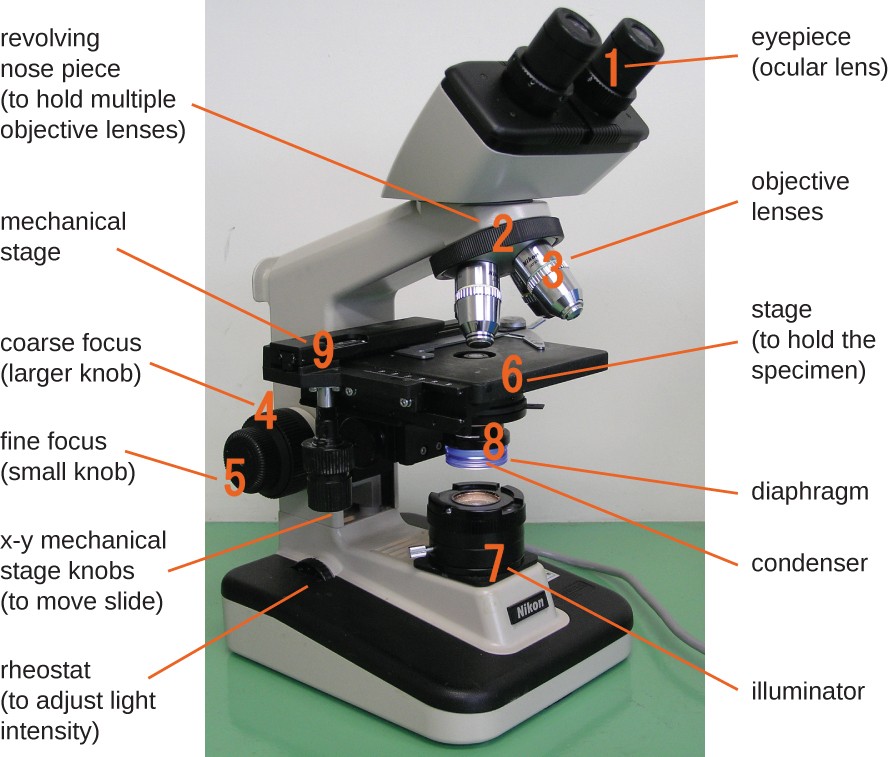
Optical electron and scanning probe microscopy along with the emerging field of X-ray microscopy. They play a major role in ensuring clear sharp images are produced with a high magnification of 400X and above. The spatial resolution that can be achieved with any light-based microscopy is however limited to about 200 nm in the imaging plane and 500 nm along the.
Specific cellular components may be observed through molecule-specific labeling and light microscopy allows the observation of structures inside a live sample in real time. Describe the ordered structures of cornea and lens that allow them to be translucent and explain how opacity can develop under pathologic conditions. In this view it is difficult to specify the edges of the cells and to interpret the cause of the dark and light areas in the image.
Tomography is imaging by sections or sectioning that uses any kind of penetrating waveThe method is used in radiology archaeology biology atmospheric science geophysics oceanography plasma physics materials science astrophysics quantum information and other areas of scienceThe word tomography is derived from Ancient Greek τόμος tomos slice. An improved image reconstruction algorithm increases time resolution and maximal imaging time for super-resolution microscopy. Super-resolution microscopy has overcome a long-held resolution barrierthe diffraction limitin light microscopy and enabled visualization of previously invisible molecular details in biological systems.
Since their conception super-resolution imaging methods have continually evolved and can now be used to image cellular structures in three dimensions. 3 cells are exposed to a significantly smaller amount of light Fig 1. Heilemann in Comprehensive Biophysics 2012 Abstract.
They are found under the stage next to the diaphragm of the microscope. Yet many students and teachers are unaware of the full range of features that are available in light microscopes.
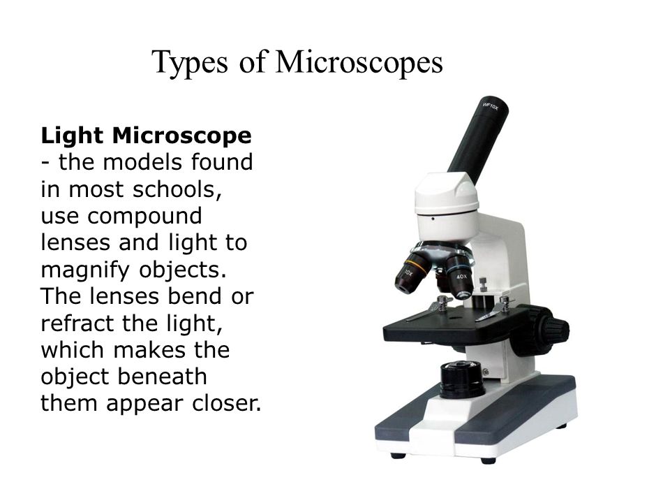
Microscopes Light Microscope The Models Found In Most Schools Use Compound Lenses And Light To Magnify Objects The Lenses Bend Or Refract The Light Ppt Download

Light Microscopes An Overview Sciencedirect Topics

Light Microscope Definition Principle Types Parts Labeled Diagram Magnification

Laboratory Equipment Light Microscope Used For Magnifying Microscopic Objects Dissecting Microscope Used To View Small Animals Plants Or Organs When Ppt Download


0 Comments